


Full-Mouth X-Ray Series (FMX)
A single image shows one area. An FMX shows the whole mouth: the root of every tooth, the bone level, and everything hidden between teeth and beneath crowns.
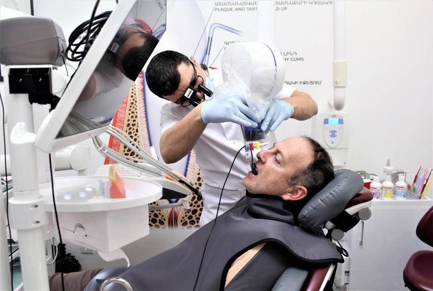
Digital sensor imaging with a protective lead apron
What an examination cannot see
Roughly 40 percent of every tooth surface is out of reach of visual examination — the contact areas between teeth, the roots, the bone. That is exactly where most decay and periodontal disease begins. Radiographs make those 40 percent visible.
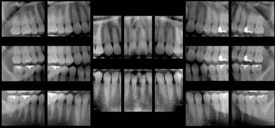
Actual FMX series, White Castle Dental Clinic, Yerevan.
What an FMX reveals
Each image covers two to three teeth including the root and surrounding bone. Together they form a map of the entire mouth.
- Hidden decay between teeth
- Bone level around every tooth
- Inflammatory lesions at the root apex
- Quality of previous root canal treatment
- Separated instruments and unfilled canals
- Decay beneath crowns and fillings
- Subgingival calculus deposits
- Retained roots and impacted teeth
- Cysts and bone changes
- Bone condition around implants
About radiation — straight answer
This is the most common question, and a fair one. Here is what the dose involves and what we do to keep it low.
- Digital sensor, not filmA digital sensor requires substantially less radiation than the older film technique, and the image appears on screen immediately.
- Lead apron with thyroid collarThe lead apron with thyroid collar shields the thyroid gland and chest. Used for every single exposure.
- Positioning holdersDedicated holders place the sensor correctly the first time. That means no repeat exposures, and therefore less radiation.
- Strict indicationsAn FMX is prescribed only where it genuinely changes the plan — comprehensive treatment planning, periodontal disease, or implant preparation.
The exact dose depends on the unit and technique. Your dentist can give you the figure for our equipment at your visit.
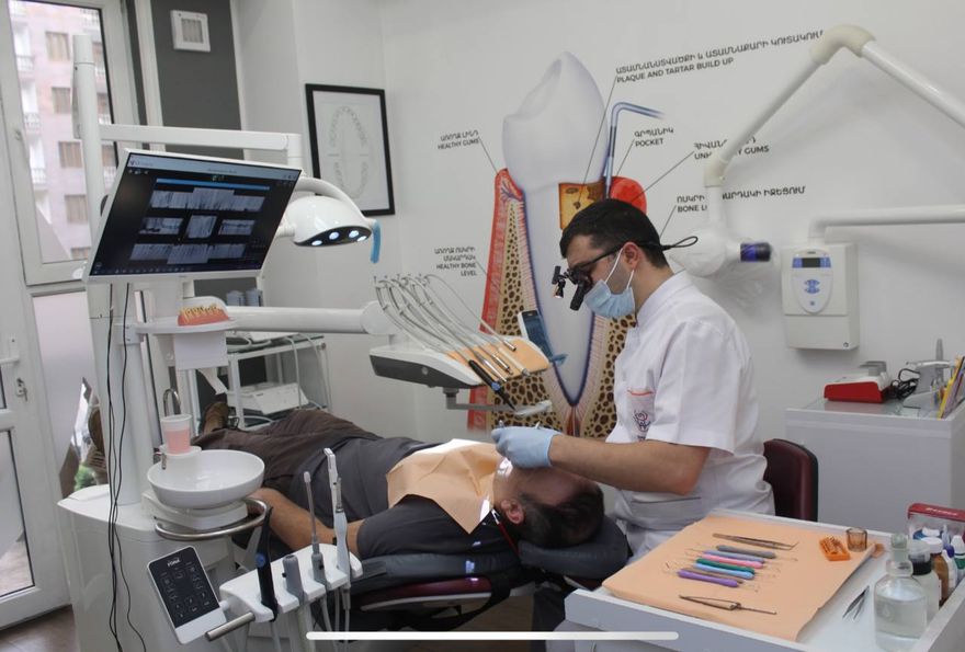
Images are reviewed immediately, with you in the room
When an FMX is needed
An FMX is not a routine check-up image. It is taken when a complete picture is required rather than a check of one area.
First comprehensive visit
When a treatment plan is being built for the whole mouth, not a single tooth.
Periodontal disease
Bone loss can only be assessed radiographically. Without it, a periodontitis diagnosis is incomplete.
Implant preparation
The condition of adjacent teeth and bone determines the plan. The FMX is supplemented by CBCT.
Before prosthetic work
Roots of abutment teeth for crowns and bridges must be verified before any work begins.
Unexplained pain
When the source of pain cannot be identified on examination.
After a long gap in care
If you have not seen a dentist in several years, one image is not enough.
Frequently asked questions
Is it safe?
The radiation dose from intraoral imaging with a digital sensor is very low. A lead apron with thyroid protection and positioning holders are used, which eliminates repeat exposures. The exact dose depends on the unit and can be confirmed at your visit.
Why is one image not enough?
A single intraoral image covers two to three teeth. Assessing the whole mouth requires 14 to 20 images from different angles. Otherwise a significant share of the problems simply falls outside the frame.
How long does it take?
15 to 20 minutes. The images appear on screen immediately and the dentist reviews them with you present.
I am pregnant — can I have it done?
Elective radiographic examination is normally postponed during pregnancy. In acute situations the decision is made individually with full protective measures in place. Always tell your dentist if you are pregnant.
How often is it repeated?
An FMX is not an annual procedure. It is repeated every few years or when the clinical situation changes substantially. Routine monitoring is done with individual targeted images.
Can I have copies of the images?
Yes. The images are digital and can be given to you or sent to another clinician.
Book a diagnostic visit
Radiographic status, microscope examination and a treatment plan with options and timeframes.
White Castle Dental Clinic · +374 12 336699 · 6/1 Nikoghayos Adonts St., Yerevan 0014, Armenia