


Detecting decay between teeth: bitewing radiographs
Around 40 percent of every tooth surface is out of reach of a visual examination — precisely where teeth touch each other. A bitewing shows that zone and finds decay while it can still be treated with a filling rather than a crown.
A bitewing captures the crowns of the upper and lower teeth at the same time, with the jaws closed. It is the only method that shows contact surfaces without distortion and lets the depth of a lesion be assessed layer by layer through enamel and dentine.
What it looks like
Both arches appear in contact. The dark areas between the teeth are zones a mirror and explorer cannot reach at all.
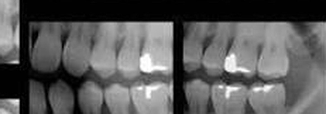
Actual bitewings from a patient at White Castle Dental Clinic, Yerevan. The bright areas are existing restorations.
What the image shows
A bitewing answers questions an examination cannot.
- Hidden decay between teethContact surfaces are the most common site of decay in adults and teenagers.
- Decay beneath old fillings and crownsSecondary decay develops along the margin of a restoration and is invisible from outside.
- Depth of the lesionIt shows whether the lesion has reached dentine. That determines whether a filling is needed or monitoring and fluoride are enough.
- Bone levelEarly signs of periodontitis in the posterior segments.
- Overhanging restoration marginsExcess material in the interdental space that traps plaque.
- Subgingival calculusCalcified deposits appear as projections on the contact surface of the root.
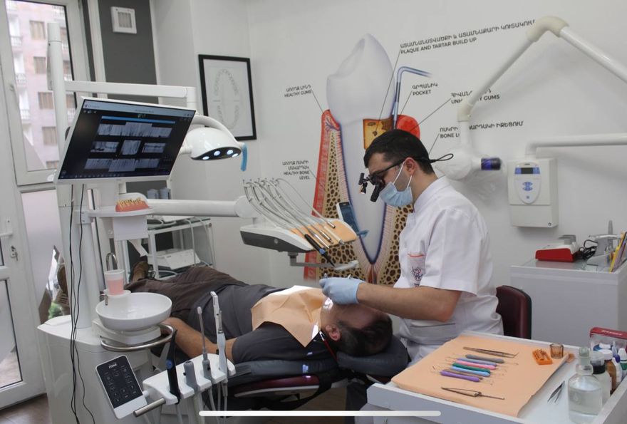
Images appear on screen immediately and are reviewed with the patient.
Bitewing, full series or CBCT — which and when
These are not interchangeable. Each answers a different question, and ordering a larger study “just in case” is the wrong instinct.
Bitewing
Contact surfaces of the posterior teeth and the bone level in those areas. Roots fall outside the frame.
Caries monitoring, routine examination
Full-mouth series (FMX)
Every tooth with its root and surrounding bone, 14–20 images.
Comprehensive planning, periodontitis, implant preparation
CBCT
A three-dimensional volume: bone, canals, sinuses, anatomical structures.
Implant surgery, complex extractions, impacted teeth
How it is taken
The accuracy of a bitewing depends entirely on sensor position. A few degrees of tilt overlaps adjacent teeth and makes the image useless — which is why we work with positioning holders rather than freehand.
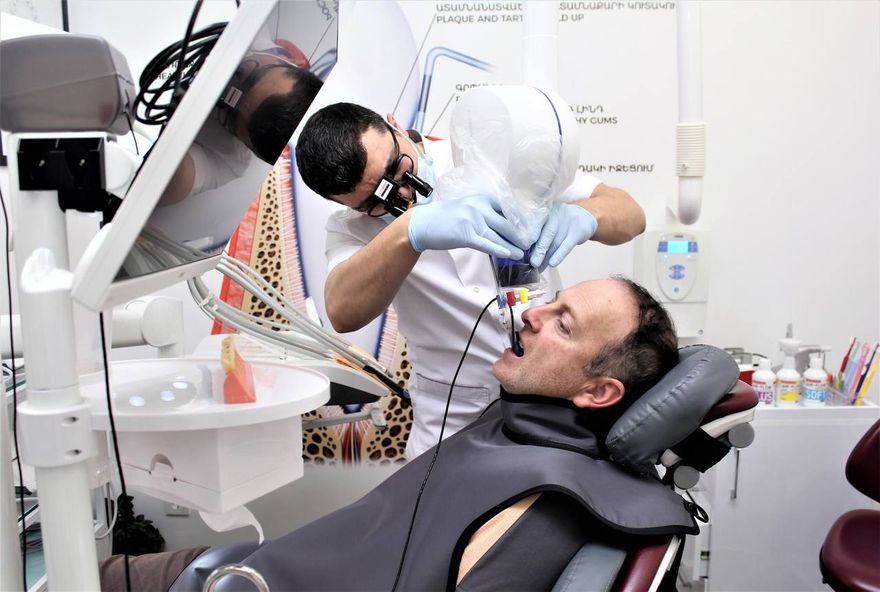
Imaging with a holder: the sensor sits parallel to the teeth and the beam is aimed through the ring.
- Digital sensorA substantially lower dose than film, with the image appearing on screen immediately.
- Holders with an aiming ringThe holder keeps the sensor strictly parallel to the teeth, and the ring sets the direction of the beam. This eliminates overlapped contacts and repeat exposures.
- Colour-coded holdersA dedicated holder for each type of projection. Picking the wrong one is almost impossible.
- Lead apron with thyroid collarUsed for every single exposure, without exception.
- Single-use barrier and autoclavingThe sensor is covered with a protective sheath, and the holders are steam sterilised after every patient.
When it is taken, and how often
The interval is set individually, by caries risk rather than by the calendar.
First examination
The baseline. Without it there is no way to tell whether a process is progressing or static.
Routine monitoring
Less often at low caries risk, more often at high risk. The decision follows the clinical picture, not a schedule.
Before prosthetic work
Abutment teeth for crowns and bridges are checked before any work starts.
Teenagers and young adults
The age of highest risk for interproximal decay.
Food packing between teeth
A frequent sign of a cavity or an overhanging restoration margin.
Follow-up after treatment
Assessment of marginal fit on completed restorations.
Frequently asked questions
Does it hurt?
No. The sensor is held by a positioner and the patient simply bites together. It can be uncomfortable with a strong gag reflex, in which case the technique can be adapted.
Why is an examination not enough?
Contact surfaces are physically out of reach of a mirror and explorer. Decay there is found on examination only once it has already broken the tooth down from the inside.
What is the radiation dose?
Imaging is done with a digital sensor, which needs substantially less radiation than film. A lead apron and positioning holders are used, which eliminates repeat exposures. Your dentist can give you the figure for our equipment at your visit.
Can it be done during pregnancy?
Elective imaging is normally postponed during pregnancy. In an acute situation the decision is made individually with full protective measures. Always tell your dentist if you are pregnant.
How many images are needed?
Usually two or four — one or two per side. That covers all the contact surfaces of the posterior teeth.
Can I have copies?
Yes. The images are digital and can be given to you or sent to another clinician.
Book a diagnostic visit
Examination under the microscope, radiographic diagnosis and a treatment plan with options and timeframes.
White Castle Dental Clinic · +374 12 336699 · 6/1 Nikoghayos Adonts St., Yerevan 0014, Armenia